


Have you ever had knee pain? Extremely uncomfortable when you suddenly start drowning or can’t get down without pain in the knee. Arthrosis of the knee joint is not life -threatening, but dramatically worsens its quality.
What is knee arthrosis?
Knee arthrosis(gonarthrosis, osteoarthritis, osteoarthritis of the knee joint). Gonarthrosis is arthrosis of the knee joint (the disease has nothing to do with gonorrhea). In further cases, surgery only helps. Do you need itThen don't let yourself be in that situation.
Causes of knee joint arthrosis.Distinguish between primary and secondary arthrosis of the knee joint. If the cause of the disease is unknown, then such arthrosis is called primary, it is inherited through the maternal line. If the grandmother suffers from arthrosis of the knee joint, then the daughter and granddaughter may develop the disease at a younger age.
Secondary arthrosis develops as a result of trauma, congenital anomalies of the knee joint, physical load (sports, occupation), endocrine disorders.
Risk factors are overweight, female sex, old age. Cartilage is very sensitive to the decline of female sex hormones, with menopause, all joints begin to "collapse". Therefore, older women who are overweight experience arthrosis of the knee joint more severely and more frequently.
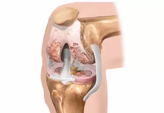
Anatomy of the knee.The knee joint is formed by the femur, tibia and patella. The articular surface of the bone is covered with a layer of cartilage. The extra cartilage spacer between the bones is called the menisci and is protective. The knee joint has the largest synovium, which forms large rotations and bursae.
The joint cavity is filled with synovial fluid, which nourishes the articular cartilage. Synovial fluid contains hyaluronic acid, which is needed to smooth the articular surface smoothly. Its ligaments, muscles and tendons guide and restrict movement in the joints.
General description.With arthrosis of the knee joint, destruction of the articular cartilage occurs. There are three stages of knee arthrosis. In the first stage, the nutrition of the articular cartilage and menisci is disrupted. Cartilage loses its elasticity and cracks. Abnormal friction occurs between bones. Joint strain is accompanied by inflammation and pain in the knee.
In the second stage, destruction of articular cartilage and menisci begins. Bone responds to load with marginal growth - osteophytes ("thorns"). The amount of intra-articular fluid decreases, the narrowing of the joint space increases. As a result, knee pain already occurs during regular exercise, walking.
In the third stage, pronounced knee joint bone deformity with significant natural movement restriction is shown.
Symptoms of arthrosis of the knee joint.The main symptoms of arthrosis are pain, limitation of movement and deformity of the knee joint. Arthrosis of the knee joint is long -term, with a slow and irreversible increase in symptoms. If the pain in the knee arises suddenly, suddenly, for the first time, it is most likely not arthrosis.
Arthrosis of the knee joint begins gradually with minor discomfort or pain in the knee during overload, long walks, when descending on the ground floor, ascending from a squatting position. At rest, the pain passes quickly.
In the second stage, knee pain is already visible with normal exercise. The volume of active movement in the knee joint decreases. The shape of the joint changes due to bone defects and abnormal fluid accumulation in the joint.
In the third stage, the pain becomes chronic, occurring not only during movement, but also at rest. Night pain interferes with sleep. The knee is difficult to install on the bed without pain. Swelling of the joints indicates an increase in inflammation. The mobility of the knee joint is reduced to a minimum.
The joints are significantly deformed, the foot becomes O or X -shaped. In severe cases, there is complete destruction of the joint with the development of ankylosis (immobility).
With knee joint arthrosis, there are 4 types of pain:
- mechanical pain arises under the influence of physical activity during the day and disappears during night rest. This knee pain is associated with a decrease in cartilage capacity and shock -absorbing structure. Knee pain is localized, as a rule, in the anterior and internal areas of the knee joint and the upper part of the lower leg.
- night pain is associated with venous blood stasis, increased intraosseous pressure in the joints and inflammation.
- "Starting" pain occurs after a period of rest, disappearing 15-20 minutes after movement in the joints. This knee pain is caused by friction of the articular surface, where fragments of cartilage damage are stored.
- persistent knee pain due to muscle spasms, as well as the development of synovitis.
Complications of knee arthrosis.Synovitis is an inflammation of the synovial membrane, which covers the articular cavity from the inside. Signs of inflammation: swelling, fever, redness, pain, joint dysfunction.
Typically, the knee joint contains 3-5 ml of synovial fluid. With disease of the joints, increased production of inflammatory fluid occurs. The volume of effusion (pathological fluid) can reach 30-70 and even more than 100 ml. The knee effusion first fills the cavity on the inside of the patella (medial fossa). With increasing volume, the upper volvulus fills, with large swelling above the patella ("horse's saddle").
Baker's cyst occurs with a marked increase in the amount of intra-articular fluid. A round elastic bulge is formed in the popliteal region. This is not a tumor, not oncology, and does not need to be operated on. Baker’s cyst can cause discomfort, pressure, and pain in the knee when moving. The diameter of the cyst is from 2 to 6 cm. With a larger size, cysts can press on the adjacent peroneal nerve with the development of weakness and numbness in the legs.
Diagnosis of arthrosis of the knee joint.Laboratory tests are not useful for diagnosis, but are used to rule out other conditions with knee pain. With arthrosis, blood volume indicators without inflammatory changes, leukocytes and ESR are within normal limits. The rheumatic test was negative. Uric acid levels are in the normal range.
X-rays show bone changes in the joints, excluding the cause of the joint pain trauma. In our country, X-ray classification of arthrosis according to stage is used.
Stage 1 - the presence of marginal bone growth with slight narrowing of the joint space;
Stage 2 - joint space is narrowed more clearly, subchondral sclerosis occurs;
Stage 3 - sharp narrowing of the joint space, leveling of the articular surface, expansion of cysts;
MRI of the knee is indicated in the early stages of the disease, when radiological changes have not yet been seen, but the patient experiences typical knee pain. With the help of MRI, you can assess the condition of cartilage, menisci, ligaments, tendons. Ultrasound of the knee joint helps visualize soft tissues (menisci, muscles, ligaments), to assess the amount of effusion.
Arthroscopy is the most accurate method for diagnosing arthrosis of the knee joint. A special probe is inserted into the joint cavity and the doctor assesses the degree of cartilage destruction under a microscope.
Treatment of knee arthrosispresent a difficult task. In each case, you should choose an individual treatment program.
When you start saying superficial things during a negotiation, the patient looks surprised at first. Is this what we want? Give me a magic injection so my knee doesn’t hurt anymore. We must clarify that there is no single method that can eliminate arthrosis. To recover, you need to move, lose weight, register to the pool. And someone wants to lie on the couch, grow a "beer belly", seize the problem with a lot of drugs and healthy. But dear !!! In this case, the drug is ineffective.
Painkillers don’t cure, they just relieve pain. Anti-inflammatory drugs are prescribed only during periods of increased pain in the knee joint. A number of non -steroidal drugs, by relieving pain, contribute to the destruction of cartilage. The healing ointment does not cure for knee arthrosis, but helps a little in relieving knee pain. With edema, redness of the joints, warming ointments and compresses are contraindicated; it is better to use local remedies with non-steroidal anti-inflammatory drugs.
Chondroprotectors do not relieve pain, are expensive, and need to be taken over a long period of time. I consider them "dummies" and practically don’t appoint them. Currently, avocado and soy extracts have appeared in pharmacies, but I have not used this drug in my clinical practice and do not have my own opinion on its effectiveness.
For the treatment and prevention of arthrosis of the knee joint, it is necessary to perform proper physiotherapy exercises in a sitting or lying position. Squatting and jumping are strictly prohibited. Cycling, swimming or working out in the water, skiing is useful. And labor exploitation in the country often causes increased knee pain. With arthrosis of the knee joint, running, brisk walking upwards, weight lifting is not recommended.
Diet for knee joint arthrosis.The knee joints bear the load in the form of their own weight. Therefore, overweight people need to lose weight at least 3-5 kg. And some patients have to lose more than a dozen kilograms. Otherwise, the treatment will not be effective. There is no need to "sit" on some kind of diet, it is dangerous for the body.
You need to change your eating habits for the rest of your life, "stop loving" all harmful products (sugary, starchy foods, beer, etc. ). Eating properly should be a habit. To lose weight, you need to eat the right foods every 3 hours.
To reduce inflammation in the joints, homeopathy recommends foods that alkalize the blood and intraarticular fluids. For this purpose, it is necessary to sharply limit meat intake, and increase the amount of vegetables and fruits in the diet.
It is believed that sausages, sausages, smoked meats, fast foods enhance the inflammatory process in the joints. Instead of pharmaceutical chondroprotectors, I recommend eating properly prepared jelly meat.
Orthopedic correction reduces stress on the knee joint. If you are experiencing pain in the knee joint, you will need to take the patella. In the case of an extension, walking with a cane is indicated. When shortening the foot, a heel insole is recommended. Recently, it has become fashionable to use kinesio cassettes. This is an adhesive tape made of natural cotton that is glued around the affected knee, not restricting its movement, but helping to relax the joint and reduce muscle spasms.
I consider interstitial electrical stimulation to be the most effective method of treating pain in arthrosis. In combination with hirudotherapy (leech therapy) and pharmacopuncture, VTES provides excellent results. I will give you a case from training.
A 54 -year -old man with stage II arthrosis of the right knee joint turned to me for help. Knee pain bothered him for 6 years. Over the years, she underwent numerous courses of drug therapy, physiotherapy, blockages with corticosteroids, and repeated courses at a rehabilitation center. But the patient's condition only got worse. He came to me for consultation for advice on whether to agree to joint replacement surgery or try something else conservatively. I didn’t have to persuade him for a long time, he immediately agreed to the treatment I suggested.
In the first session, I gave him 6 leeches, which helped relieve joint swelling and relieve night pain. The knees have become easier and free to move. The man felt a little relieved. Then we performed 3 interstitial electrical stimulation procedures and almost completely stopped the pain syndrome.
Subsequently, success was combined with the introduction of homeopathic preparations with anti-inflammatory and chondroprotective effects to acupuncture points. After 3 weeks from the start of therapy, the patient discarded the cane and began to move freely, without suffocation. 3 years have passed since then. Knee pain does not return. Once a year, we run a VTES session with the goal of prevention.
Intra-articular injections with hormones are very effective in emergencies to relieve severe pain, swelling and inflammation. The indication is effusion, it is forbidden to make blockages with corticosteroids on "dry joints"! They temporarily relieve the pain, but such injections do not cure the arthrosis itself, and the cartilage is subsequently destroyed even more. It should be performed by a trained physician who knows the indications, contraindications, medications, point of administration. In total, no more than 3 blocks are required for each connection.
After eliminating the swelling and inflammation, a hyaluronic acid preparation, called a liquid prosthesis, is injected into the joint. They act on the joints as a natural lubricant, improve the sliding of the bone surface, and restore the shock -absorbing function of the cartilage. But hyaluronic acid preparations are expensive, and they only last 6-8 months. There is no point in giving hyaluronic acid preparations with complete loss of joint space and in patients over 65 years of age.
Treatment with folk remedies.You can use tincture or decoction of cinquefoil, compresses with carrots, radish or ginger, turpentine bath.
Joint endoprosthetics can only be performed in the event of serious knee joint dysfunction, because after 10-15 years these joints should be changed again. Is there enough strength and health every 10-15 years for surgery under general anesthesia and subsequent recovery??? So do not rush to agree to the operation! Take care of your joints!